Microorganisms all over our body
We’re never alone, and that’s a good thing. Many different microorganisms including bacteria, viruses and fungi live on and inside our bodies. These microorganisms are called the microbiome. Almost all parts of our body, such as the gastrointestinal tract, host their own specific microbiome which fulfils important functions. In our guts for example, the microbiome can assist in digestion and help us absorb nutrients. The organisms that make up the microbiomes are as diverse as the different cells in our body and can actively impact our health. A complete lack of beneficial microbiome components, presence of harmful microorganisms or an imbalance of specific species can negatively impact our general health. The goal of the HoloGen project is to understand how these microbial communities interact with their host, which can be us humans or any other animal, and how those interactions shape overall health.
Shining light on small interactions
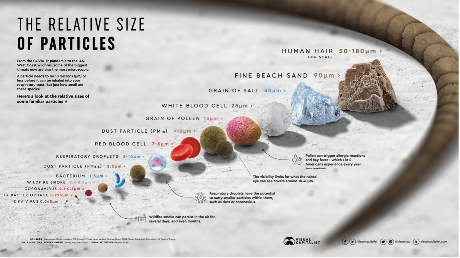
As you can see in this infographic obtained from Visual Capitalist, microorganisms are so small they can’t be seen by the naked eye like our hair or a grain of salt. For looking at such tiny cells, a microscope is needed. But even under the microscope many microorganisms look almost identical, therefore we need to develop ways to distinguish them.
My goal during the PhD is to establish a new way to visualise the microbes and their interaction inside the gut. In principle, the idea is to make different microbial species light up in different colours to identify them. In one sentence, I will design so-called probes containing a fluorophore which will be specifically targeting individual microorganisms and light them up differently.
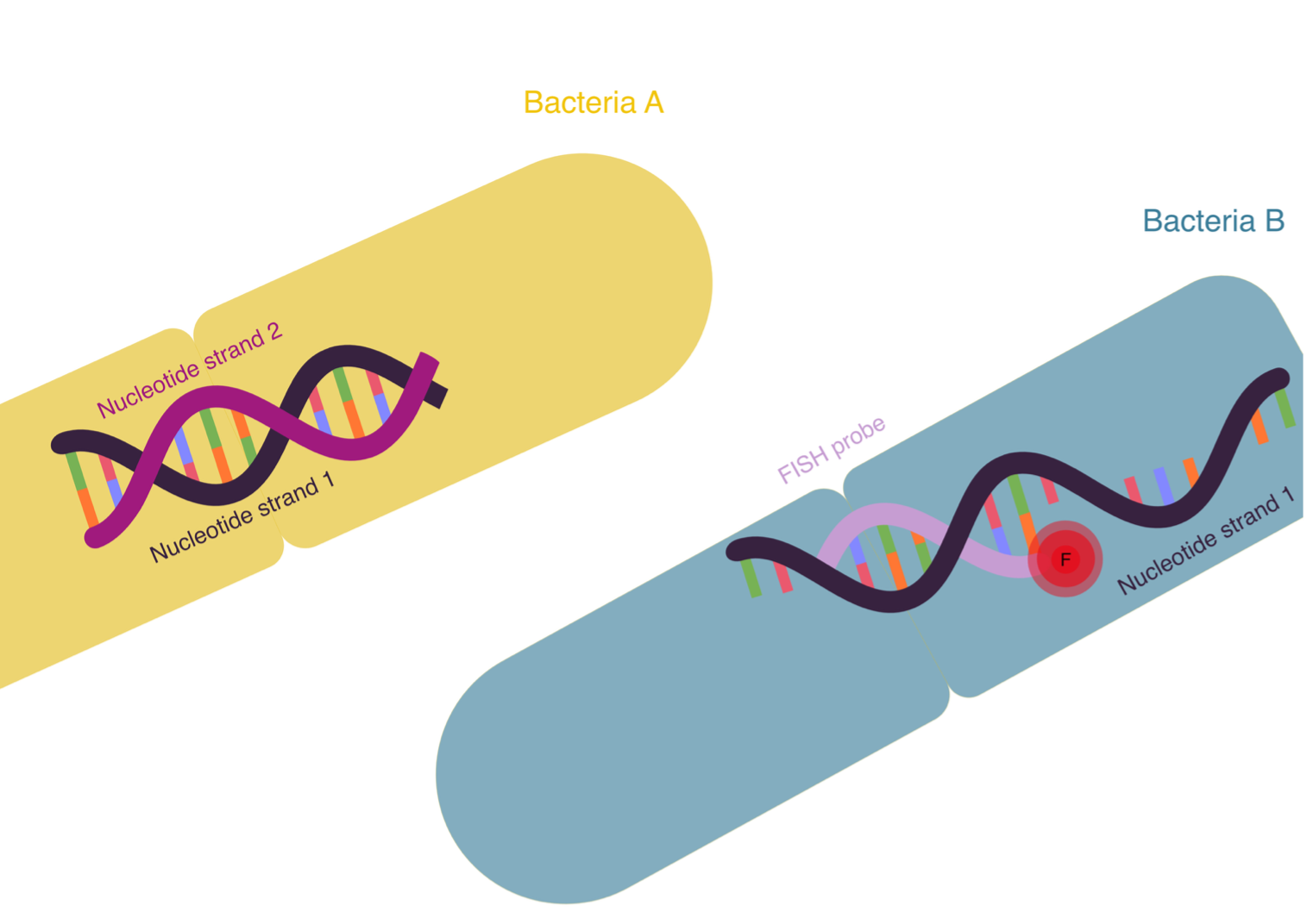
Probes are specific to the microbes or the hosts intestinal cells by targeting their respective genome. The genome consists of DNA, which is made up of two complementary strands of nucleotide sequences. This sequence is specific for each living organism and can therefore be used to tell them apart. DNA can be found in cells as on the left-hand side of the Figure in Bacteria A, with the two complementary oligonucleotide strands bound together. Sometimes, DNA also occurs as on the right-hand side of the Figure in Bacteria B, as so-called single stranded DNA. In Bacteria B it is shown that when single-stranded DNA is present, a probe that is complementary to a specific part of the sequence can bind to it. The fluorescent tag which is bound to the probe is visualised under the microscope and can be imaged, just like taking a picture of a firefly. This specific microscopic imaging approach is known as FISH (fluorescence in-situ hybridisation).
The tricky part is designing each probe in a way that it will only bind to a specific part of the genome of the microorganism I want to light up. I use computational tools to scan the genetic information of all microbiome components and identify short DNA sequences that are unique to each species. These sequences will become the targets of the probes. Once I have the FISH probes designed for all the microbiome components as well as the host intestinal cells, I can start imaging actual biological samples. With the help of veterinarian collaborators, which provide me with gut samples of agricultural animals, I hope to study the interaction of the microbiome with its host using my colourful fluorescent probes.
Why bother with this?
Currently, most microbiome studies focus on which microorganisms are present in a microbiome and how much of each species there is. But the crucial piece missing in these studies is identifying where exactly each species is located within the sample.
There’s a high chance that the positioning of the microorganisms within the microbiome is not random but is in fact meaningful. Microorganisms can be up to 1’000 times smaller than the host cells they interact with. The signals that these very small organisms produce, are also very small chemical messages and can therefore have limited range. That means, microbes tend to only influence their immediate surroundings and don’t have very far reach. So, if two microorganisms within the microbiome are located far apart, they most likely don’t interact with each other. The same applies to microbes and their potential interaction with host cells. The distance between two individual cells can determine whether they interact or not.
By investigating the spatial arrangement of microbiome components, I want to understand these local interactions. I aim to see which microbes cluster together, which locate near the host tissue, and which seem to be isolated? At the end of my PhD, I want to use these colourful maps of the microbiome to help us understand how microbial communities are organized. Through this I aim to understand how the microbiome interacts with the host, and ultimately, how these interactions can affect our health. Because sometimes, to understand life at its smallest scale, you just need to shine a little light on it.
